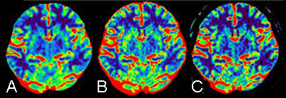
Due to the greater availability of CT/MRI perfusion imaging, it is widely accepted as a diagnostic tool for cerebrovascular disorders. Although the deconvolution method is generally employed for analysis, there is significant variation in the actual implementation of the deconvolution algorithm among analysis environments.
An identical set of the entire currently available CT/MRI perfusion data, which were obtained from different scanners in several institutes, was fed into the analyzing systems. We found that there was a substantial variation and discrepancy in the output [1,2]. Hence, it is necessary to find an optimal algorithm that produces an accurate and reproducible output.
 The mean transit time (MTT) maps from an identical CT perfusion dataset that are computed by different workstations supplied by five vendors (A-E). There is substantial variation in the appearance of the maps; these variations include the definition of cerebral ischemia in the area of the left middle cerebral artery.


